

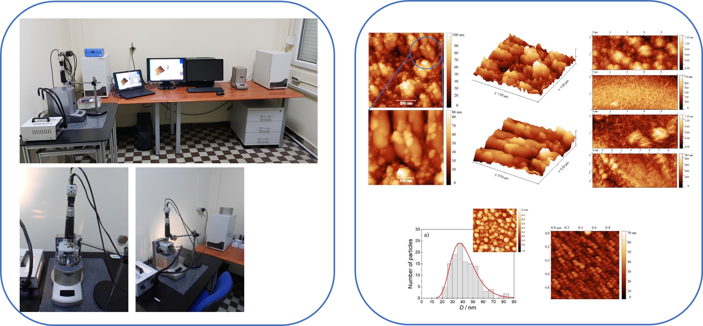
The laboratory for nanomaterials is equipped with:
Schimadzu SPM-9700HT - modes of operation:
✔ contact mode
✔ Lateral Force Microscopy, LFM
✔ Force Modulation Microscopy, FMM
✔ intermittent contact mode (TAP-AFM)
✔ Phase modulation mode
✔ Piezoresponse Force Microscopy, PFM
Schimadzu SPM-9600 - mode of operation:
✔ Scanning Tunneling Microscopy, STM.
Available scanners:
i. mid-range scanner (30 mm x 30 mm x 5 mm)
ii. HT scanner (10 mm x 10 mm x 1 mm).
In addition, the Laboratory for nanomaterials has access to the following instruments available at the Institute of Chemistry:
Through collaboration with the York-JEOL Nanocentre, York University, UK, the Laboratory for nanomaterials has also access to state-of-the-art electron microscopes, PL and XPS spectroscopy techniques.
RESEARCH
SELECTED PUBLICATIONS:
1. B. Pejova, A. Eid, L. Lari, A. Althumali, L. Šiller, A. Kerrigan, Lj. Pejov and Vlado K. Lazarov, 3D self-assembled polar vs. non-polar NiO nanoparticles nanoengineered from turbostratic Ni3(OH)4(NO3)2 and ordered β-Ni(OH)2 intermediates, Nanoscale, accepted.
2. B. Pejova, S. Premcheska, E. Sherif Miftar, From self-affine Ag to mounded Ag@Ag2O core-shell nanoplasmonic surfaces by sonochemistry, J. Phys. Chem. C 127 (2023) 11204–11217.
3. B. Pejova, E. Sherif, M. W. Minde, Sonochemically synthesized quantum nanocrystals of cubic CuInS2: Evidence for multifractal surface morphology, size-dependent structure, and particle size distribution, J. Phys. Chem. C, 124 (2020) 20240-20255.
3. T. Zibar Belasic, B. Pejova, H. Otmacic Curkovic, E. Kamenar, B. Cetenovic, S. Spalj, Influence of Intraoral Application of Antiseptics and Fluorides During Orthodontic Treatment on Corrosion and Mechanical Characteristics of Nickel-Titanium Alloy in Orthodontic Appliances, The Angle Orthodontist, 91 (2021) 528-537.
4. B. Pejova, Ultrasound driven mound to self-affine surface structure transition in nanostructured films: one-pot route to cubic CuInS2 quantum dots with controlled structure and tunable band gap energy, Journal of Materials Science: Materials in Electronics, 30 (2019) 12679-12694.
5. I. Musa Trolic, Z. Todoric, D. Pop Acev, P. Makreski, B. Pejova, S. Spalj, Effects of the presence of probiotic bacteria in the aging medium on the surface roughness and chemical composition of two dental alloys, Microscopy Research & Technique, 82 (2019) 1384-1391.
6. B. Pejova, Optical absorption of semiconductor quantum dot solids, Semiconductor Science and Technology 29 (2014) 045007 (15 pages) (Paper selected for inclusion in IOPselect, on the basis of the following criteria: i) substantial advances or significant breakthroughs; ii) a high degree of novelty; iii) significant impact on future research; http://Select.iop.org.).
7. B. Pejova, Optical phonons in nanostructured thin films composed by zincblende zinc selenide quantum dots in strong size-quantization regime: competition between phonon confinement and strain-related effects, Journal of Solid State Chemistry 213 (2014) 22-31.
8. B. Pejova, Three-dimensional assemblies built up by quantum dots in size-quantization regime: Band gap shifts due to size-distribution of cadmium selenide nanoparticles, Journal of Solid State Chemistry 207 (2013) 147-151.
9. B. Pejova, Phonon confinement and related effects in 3D assemblies of cubic cadmium selenide quantum dots synthesized by conventional chemical and sonochemical routes, Journal of Physical Chemistry C 117 (2013) 19689-19700.
10. B. Pejova, I. Bineva, Sonochemically synthesized 3D assemblies of close-packed In2S3 quantum dots: Structure, size dependent optical and electrical properties, Journal of Physical Chemistry C 117 (2013) 7303-7314.
11. J. Bloch, O. Levy, B. Pejova, J. Jacob, S. Curtarolo, B. Hjörvarsson, Prediction and hydrogen acceleration of ordering in iron-vanadium alloys, Physical Review Letters 108 (2012) 215503 (5 pages).
12. B. Pejova, B. Abay, Nanostructured CdSe films in low size-quantization regime: tem-perature dependence of the band gap energy and sub-band gap absorption tails, Journal of Physical Chemistry C 115 (2011) 23241-23255.
13. B. Pejova, D. Nesheva, Z. Aneva, A. Petrova, Photoconductivity and relaxation dynamics in sonochemically synthesized assemblies of AgBiS2 quantum dots, Journal of Physical Chemistry C 115 (2011) 37-46.
14. J. Bloch, B. Pejova, J. Jacob, B. Hjörvarsson, Hydrogen-vanadium system in thin films: Effect of film thickness, Physical Review B 82 (2010) 245428.
15. B. Pejova, B. Abay, I. Bineva, Temperature dependence of band gap energy and sub-band gap absorption tails in strongly quantized ZnSe nanocrystals deposited as thin films, Journal of Physical Chemistry C 114 (2010) 15280-12291.
16. B. Pejova, The higher excited electronic states and spin-orbit splitting of the valence band in three-dimensional assemblies of close-packed ZnSe and CdSe quantum dots in thin film form, Journal of Solid State Chemistry, 181 (2008) 1961-1969.
17. B. Pejova, I. Grozdanov, D. Nesheva, A. Petrova, Size-dependent properties of sonochemically synthesized three-dimensional arrays of close-packed semiconducting AgBiS2 quantum dots, Chemistry of Materials 20 (2008) 2551-2565.
18. B. Pejova, A. Tanuševski, A study of photophysics, photoelectrical properties and photoconductivity relaxation dynamics in the case of nanocrystalline tin(II) selenide thin films, Journal of Physical Chemistry C 112 (2008) 3525-3537.
COLLABORATION
Contact: prof. Biljana Pejova
biljana@pmf.ukim.mk